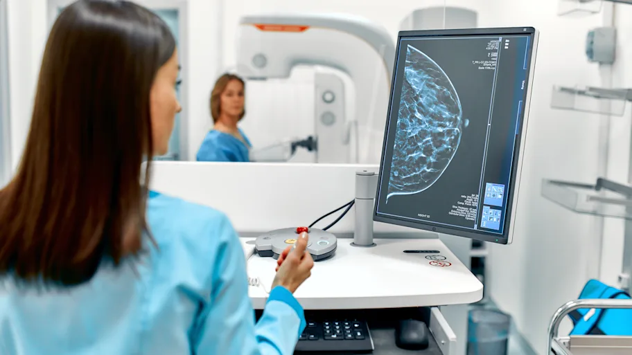
Breast cancer remains one of the most frequently diagnosed oncological conditions affecting populations globally, developing when cellular mutations occur within the breast tissue. These genetic alterations lead to uncontrolled cellular proliferation, eventually forming a malignant tumor that can infiltrate surrounding structures and spread to distant organs. Over the past few decades, the medical community has achieved remarkable strides in both the detection and therapeutic management of this complex disease. Today, a highly integrated, multidisciplinary approach ensures that individuals diagnosed with Breast Cancer receive individualized treatment pathways. By shifting away from a one-size-fits-all model toward precision medicine, modern oncology maximizes survival rates while aggressively protecting the patient’s long-term quality of life.
The Biological Framework and Cellular Subtypes
To effectively eradicate the malignancy, oncologists must first precisely identify the anatomical and cellular origin of the tumor. The most prevalent subtype is invasive ductal carcinoma, a cancer that originates in the milk ducts before breaching the ductal walls to invade surrounding adipose tissue. Another primary variant, invasive lobular carcinoma, begins within the lobules the specific anatomical glands responsible for producing milk.
Beyond tracking the physical origins, contemporary pathology classifies these tumors based on their molecular receptor status. Tissue samples from biopsies are rigorously tested for the presence of estrogen receptors, progesterone receptors, and the human epidermal growth factor receptor 2 (HER2). Furthermore, approximately ten to fifteen percent of diagnoses present as triple-negative breast cancer. This aggressive subtype lacks all three primary receptors, rendering traditional hormone-blocking and HER2-targeted therapies ineffective. Identifying these exact molecular and genetic signatures is an absolute prerequisite for formulating an effective, customized systemic strategy.
Advanced Diagnostics and Genomic Profiling
Early detection remains the most powerful variable in the oncological arsenal. Standardized screening protocols, primarily utilizing digital mammography, allow radiologists to detect microcalcifications and architectural distortions years before a physical lump becomes palpable to the patient. When initial screenings reveal anomalies, medical professionals deploy a secondary tier of high-resolution imaging modalities, including breast magnetic resonance imaging (MRI) and targeted ultrasonography.
These advanced imaging techniques provide a detailed, three-dimensional mapping of the breast tissue, enabling physicians to perform highly accurate, image-guided core needle biopsies. Elite medical institutions, such as Liv Hospital, integrate these sophisticated diagnostic technologies into rapid-response workflows. This immediate access to high-fidelity imaging and pathology helps secure rapid, definitive diagnoses, thereby eliminating prolonged periods of patient uncertainty and expediting the transition into active medical intervention.
Precision in Surgical Interventions
Surgical excision frequently forms the cornerstone of the primary treatment protocol. The modern surgical doctrine strongly emphasizes breast conservation therapies whenever medically and anatomically viable. A lumpectomy, occasionally referred to as a partial mastectomy, involves the precise removal of the malignant tumor alongside a clean margin of healthy surrounding tissue. This technique actively preserves the structural integrity and aesthetic contour of the breast.
For patients presenting with advanced, multifocal, or highly aggressive malignancies, a total mastectomy may be necessitated. In parallel to addressing the primary tumor, surgeons routinely perform axillary lymph node evaluations. By utilizing the sentinel lymph node biopsy technique, surgeons can inject a specialized dye or radioactive tracer to identify and remove the first nodes into which the tumor drains. This crucial evaluation dictates whether the malignant cells have migrated into the lymphatic system, a factor that heavily influences subsequent systemic treatments.
Systemic Therapies and Targeted Oncology
Following surgical intervention or occasionally preceding it in the form of neoadjuvant therapy medical teams employ a broad spectrum of systemic treatments to eradicate microscopic disease and drastically lower the risk of recurrence. Traditional cytotoxic chemotherapy circulates throughout the entire vascular system, attacking and destroying rapidly dividing cells.
However, the most profound advancements in recent years involve the rapid evolution of targeted therapies and immunotherapies. For tumors overexpressing the HER2 protein, specialized monoclonal antibodies are deployed to actively hunt down the specific cellular receptors driving the growth, effectively shutting down the tumor’s replication machinery with minimal collateral damage to healthy tissues. Similarly, oral endocrine therapies are utilized for hormone-receptor-positive malignancies. In cases where the patient harbors genetic mutations, such as BRCA1 or BRCA2, oncologists may integrate PARP inhibitors into the regimen. These specific medications prevent cancer cells from repairing their damaged DNA, ultimately causing the tumor cells to undergo programmed cell death.
Radiation Mechanics and Tissue Preservation
Radiation oncology frequently complements surgical and systemic treatments, serving as a critical safety net following breast-conserving surgeries. High-energy, precisely targeted ionizing radiation is directed at the surgical bed and surrounding tissues to completely destroy any residual, microscopic cancer cells that evaded the surgeon’s scalpel. Advanced delivery mechanisms now allow radiation oncologists to perfectly modulate the intensity of the beams, contouring the radiation field to the exact three-dimensional shape of the target area. This precision ensures that adjacent vital organs, such as the heart and the lungs, are spared from unnecessary radiation exposure.
Ongoing Surveillance and Survivorship Protocols
Transitioning from active oncological intervention to a state of long-term monitoring requires a highly structured, multidisciplinary protocol. Patients enter a rigorous surveillance phase that typically includes routine physical examinations, biannual or annual mammography, and comprehensive metabolic panels. Oncology teams focus heavily on anticipating and mitigating the late-stage side effects of aggressive treatments.
Lymphedema, a potential swelling of the arm directly related to the previous removal of lymph nodes, is proactively managed through specialized physical therapy, manual drainage, and compression techniques. Furthermore, survivorship programs emphasize the importance of ongoing cardiovascular health and bone density preservation, particularly for individuals who received anthracycline-based chemotherapies or extended endocrine suppression. This proactive vigilance ensures that physiological recovery aligns perfectly with long-term functional stability and holistic health.